Abstract
The field of imaging technology is continuously evolving, offering new possibilities for the examination, conservation and documentation of wall paintings. This paper presents a range of common imaging techniques, from in-situ microscopy to three-dimensional (3D) modelling and geospatial mapping, alongside examples of their application. By integrating relevant imaging techniques into their practice, conservators can enhance their ability to document, analyse and monitor wall paintings, and communicate those findings to others.
Introduction
The advancement of imaging techniques has significantly improved the methods available for analysing and preserving cultural heritage. This paper outlines several key imaging technologies, and their applications in wall painting conservation. The techniques are presented from micro-scale to broader imaging methods, demonstrating how each can contribute to a comprehensive conservation strategy or question. The reader is encouraged to research the specific ‘how to’ themselves.
In-situ microscopy
In-situ microscopy is a well-established technique that allows for immediate, on-site analysis without the need to remove materials for examination in the lab. A portable digital microscope, such as a Dinolite (other digital microscopes are available), connected to a laptop, provides high-resolution images of surface details; newer models of microscope may have an integrated screen for easier in-situ use. Different models of portable microscope have different camera specifications, magnification ranges and light sources. Higher camera resolution is often more practically useful than higher magnification. The best light source depends on the question being investigated. For example, ultraviolet (UV) radiation in combination with in-situ microscopy enhances the ability to examine varnishes and repaint. The UV can be integral to the portable microscope or from a secondary source such as a UV torch. Secondary visible light sources can also be used to create a raking light, which can reveal topographic variations on the micro scale.
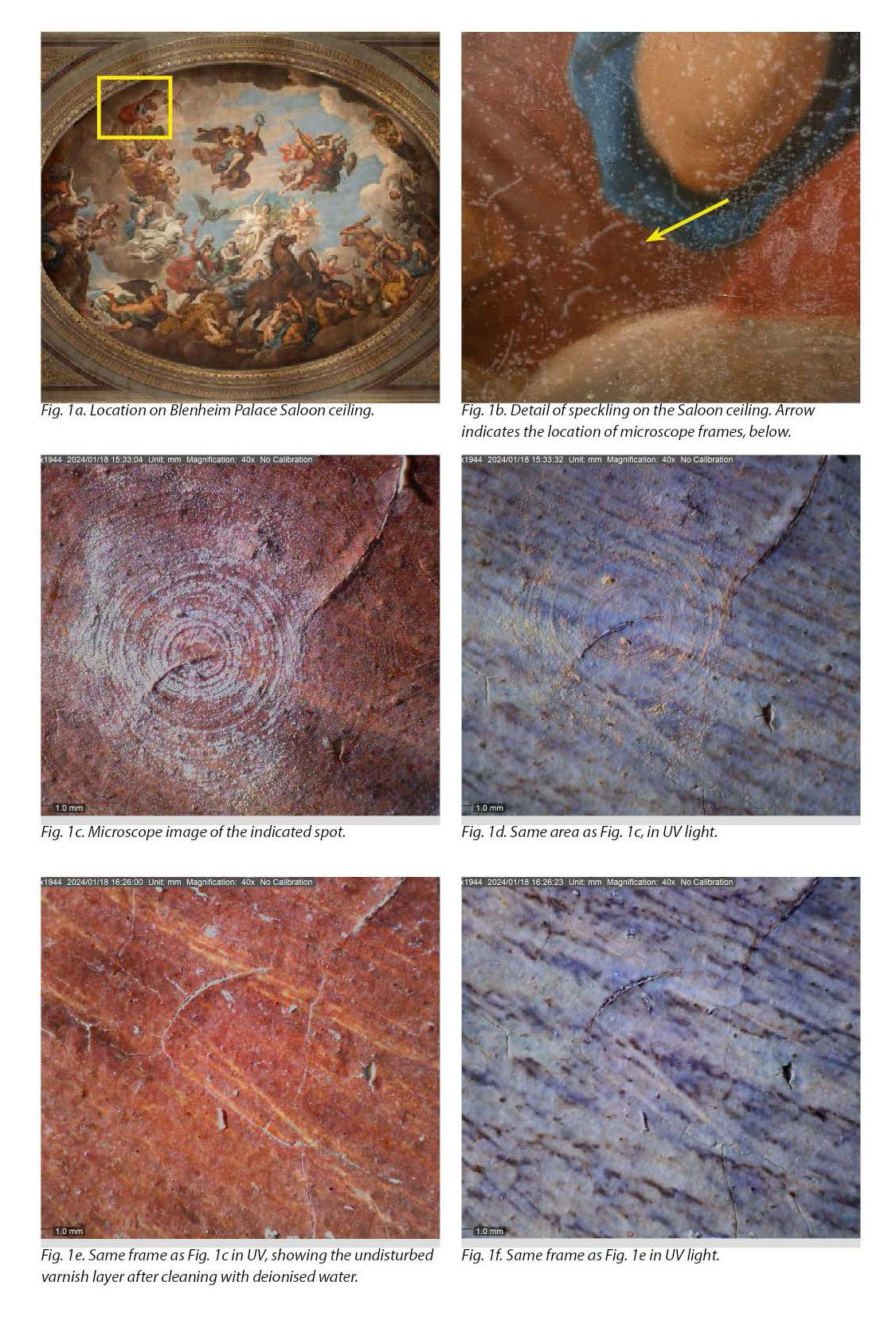
The different uses a conservator might find for in-situ microscopy include recording observations about original technology, diagnosis of deterioration phenomena and assessment of conservation interventions. For example, in Figure 1, microscope imaging reveals the cause of surface speckling (as observed with the naked eye, Figure 1b) to be condensation (see Figures 1c-1d). The same imaging technique showed that the condensation spots could be removed with water without surface damage (see Figures1e-1f). Reassessment of the exact same area – before and after conservation – was made possible by the use of topographical markers on the paint layers.
Bandpass multi-spectral imaging
Multi-spectral imaging (MSI) has been widely adopted in conservation, providing valuable insights into an artwork’s execution and material composition.1 In laboratory settings, this technique has evolved into hyperspectral imaging (HSI), generating large datasets for detailed analysis. However, on-site, broad bandpass multi-spectral imaging – using bandpass filters to capture photographs while illuminating the painting with infrared (IR) or ultraviolet (UV) radiation sources – usually provides a useful level of information to the wall painting conservator.
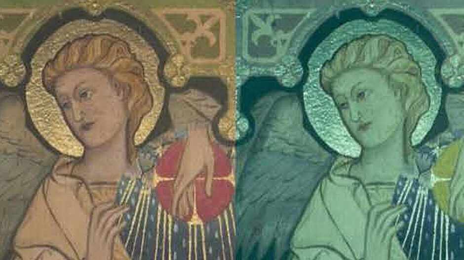
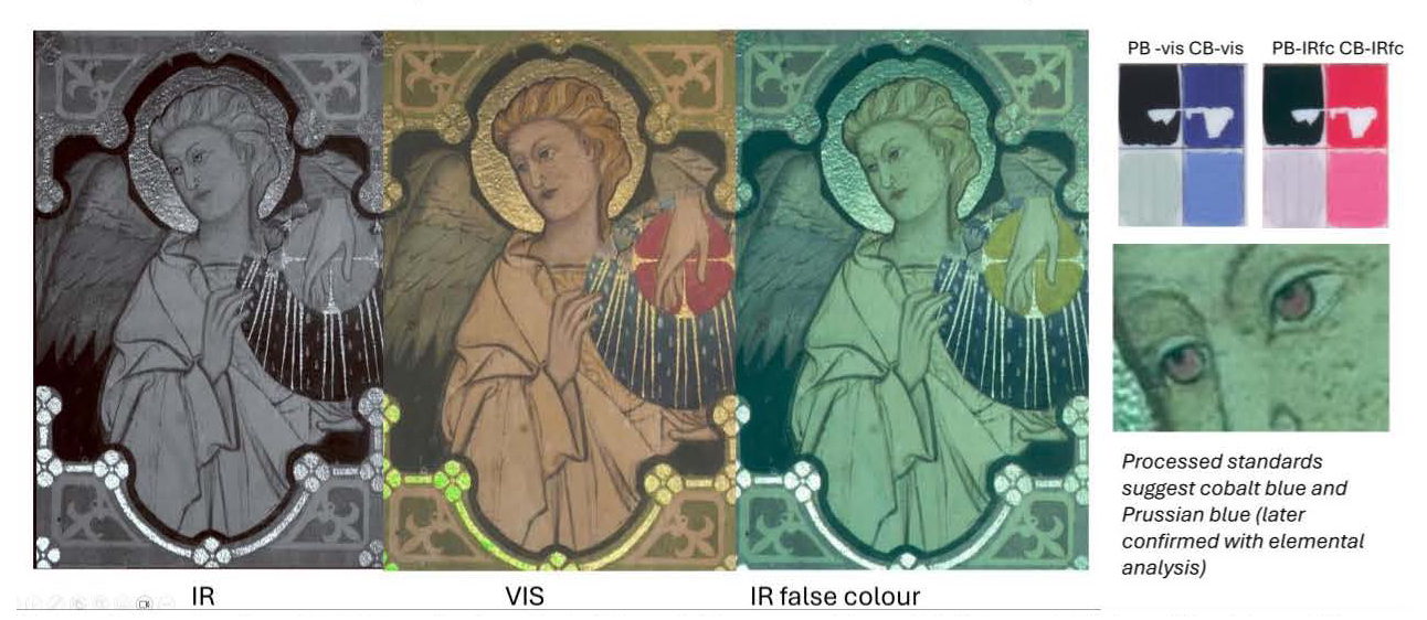
Infrared reflected (IRR) photographs can reveal underlying drawings and composite infrared false-colour images (IR-FC) – when compared to known references – can be used to characterise pigments. This technique assists in preliminary assessments and guides further analytical procedures, such as invasive sampling. In the example shown in Figure 2, the IRR image shows the preliminary pencil sketch. The IR-FC image demonstrates that at least two blues were used in the painting of this angel figure, the blue of the eyes (red in IR-FC) and the blue of the wings and orb (dark blue in IR-FC). These two blues could be characterised by comparing the IR-FC image to an IR-FC image of known pigment standards, and then firmly identified with further elemental analysis.
UV-induced visible luminescence
Ultraviolet-induced visible luminescence (UVL) photography is particularly effective in detecting varnishes and overpainting. Traditionally this has been done at close range, but now long-throw UV torches are available, UVL imaging can be carried out from significant distances (more than 20m, see Figures 3a to 3c). This capability is invaluable for the preliminary examination of wall paintings that are not immediately accessible, thereby improving planning and cost estimation processes.
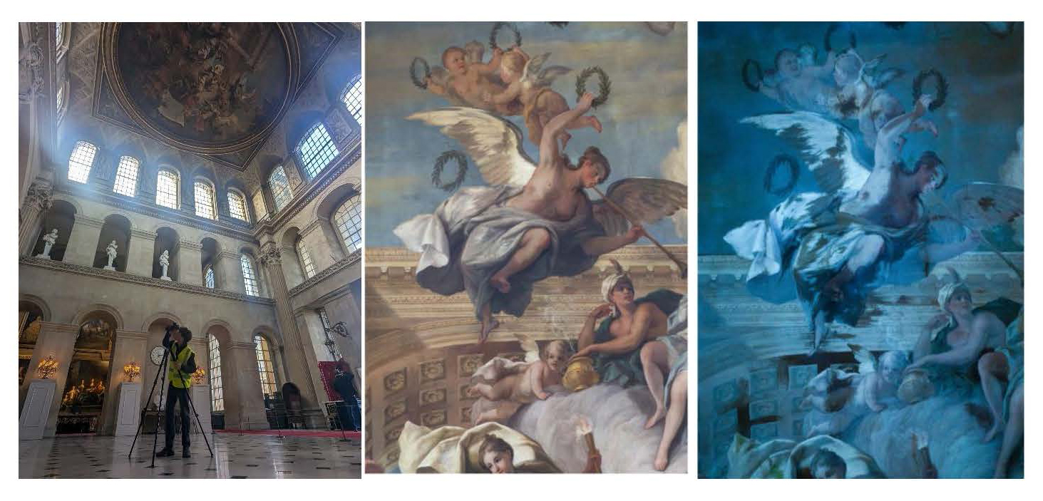
Image alignment
Presenting MSI, and generating false-colour images, depends on accurate alignment of images. Identically aligned frames are also helpful for showing comparative conservation images in reports. Software tools, such as Adobe Photoshop, provide efficient alignment functions that facilitate the visualisation of changes over time.
High-resolution imaging
High-resolution imaging, achieved by merging multiple frames into a single image, compensates for the resolution limitations of single-frame capture. This approach is particularly useful when working with large-scale paintings or artworks behind protective glass, where reflections and distortions may obscure details. High-resolution images can be produced using various software. Photoshop usually produces good merges with flat wall paintings, and surface glare can be mitigated. However, it struggles with three-dimensional features.
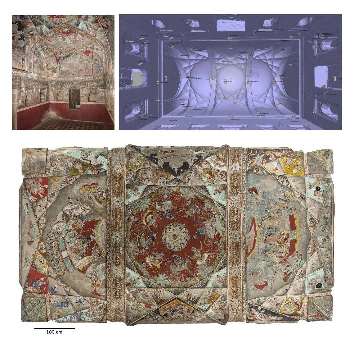
Structure-from-Motion (SfM) and 3D Modelling
For complex surface topography and three-dimensional spaces, Structure-from-Motion (SfM) software enables the creation of accurate 3D models using two-dimensional photographs. From these 3D models, orthophotos can be exported. These have many advantages over regular images or even rectified ones; they are geometrically correct, and maintain accuracy and scale across large surfaces and complex shapes. This allows for removal of line-of-sight obstructions and visualisation of architecturally complex geometry that would not be possible from a single or even multiple points of capture. For large or complex wall painting projects, orthophotos can also be used as accurate basemaps on which to record or monitor change.
Mobile applications for 3D documentation
SfM software can be processor-heavy and expensive but, inexorably, technological advancements have made 3D modelling more accessible through mobile applications such as Polycam. These applications generate 3D models directly from smartphone images, offering a cost-effective and efficient means of capturing high-quality documentation. This method of capture was recently employed by the authors in the rapid large-scale documentation of an extensive ruined heritage site with widespread wall painting remains.
GIS integration and digital archives
The integration of imaging and survey data into GIS platforms allows for spatial organisation and interactive data visualisation of conservation documentation. For instance, using GIS to map wall painting across large heritage sites or geographical areas aids in conservation management and helps strategise conservation efforts. This also improves accessibility to documentation for both conservation planning and interpretation purposes. The overlay of analytical data, such as images of paint sample cross-sections, enhances communication of findings, allows others to interrogate the data, and contributes to the comprehensiveness of digital archives.
Future directions in conservation imaging
As imaging technologies continue to evolve, their applications in conservation must be carefully considered to ensure long-term usability. The transition towards digital files and 3D models necessitates the development of robust archival strategies to preserve conservation records for future generations. Efforts should focus on standardising data formats and ensuring compatibility with emerging technologies to facilitate seamless access and analysis by future conservators. And as conservators, we must comply with record format guidance issued by clients and archives.
Conclusion
The rapid advancement of imaging techniques expands the possibilities for wall painting conservation. From in-situ microscopy to 3D modelling, these technologies provide conservators with powerful tools for analysis, monitoring, and documentation. Moving forward, it is essential to embrace these innovations, selecting what is useful to us, while ensuring the sustainability and accessibility of digital conservation records. In so doing, we will be better able to optimise our efforts as conservators, and to produce work that is intelligible to our clients and colleagues, now and into the future.
1 See, for example, the many publications of Alfredo Aldrovandi, Antonino Cosentino and Giovanni Verri, in particular: Alfredo Aldrovandi, Ottaviano Caruso, and Paola Ilaria Mariotti, “Caratterizzazione dei materiali pittorici nelle pitture murali mediante tecniche fotografiche,” OPD restauro 22 (2010): 55-80; Antonino Cosentino, “Panoramic, macro and micro multispectral imaging: an affordable system for mapping pigments on artworks,” Journal of Conservation and Museum Studies 13, no. 1 (2015); and Giovanni Verri, “The spatially resolved characterisation of Egyptian blue, Han blue and Han purple by photo-induced luminescence digital imaging,” Analytical and Bioanalytical Chemistry 394, no. 4 (2009): 1011-1021.